Being told you have one is not the same as needing surgery
Aneurysms are increasingly found on scans done for something else entirely — headaches, dizziness, a check-up. Most small aneurysms never bleed. Treating every one that is discovered would cause more harm than it prevents, because every treatment carries its own risk, and that risk is taken on a day when the patient is otherwise well.
So the first task is not technical. It is to work out, honestly, whether the aneurysm in front of us is more dangerous left alone than treated.
What we weigh
- Size and shape. Larger aneurysms and those with an irregular outline or a daughter sac carry more risk than small, smooth ones.
- Where it sits. Some locations bleed more readily than others, and some are far easier to reach safely.
- Whether it is growing. An aneurysm that has enlarged between two scans behaves differently from one that has been stable for years.
- Your age and general health, because the risk of rupture accumulates over the years you have ahead.
- Blood pressure and smoking, both of which raise rupture risk and both of which can be changed.
- Family history. An aneurysm or subarachnoid haemorrhage in a first-degree relative matters.
For many patients the conclusion is surveillance: a repeat scan at a defined interval, blood pressure treated properly, and smoking stopped. That is an active decision, not a refusal to treat.
How they are investigated
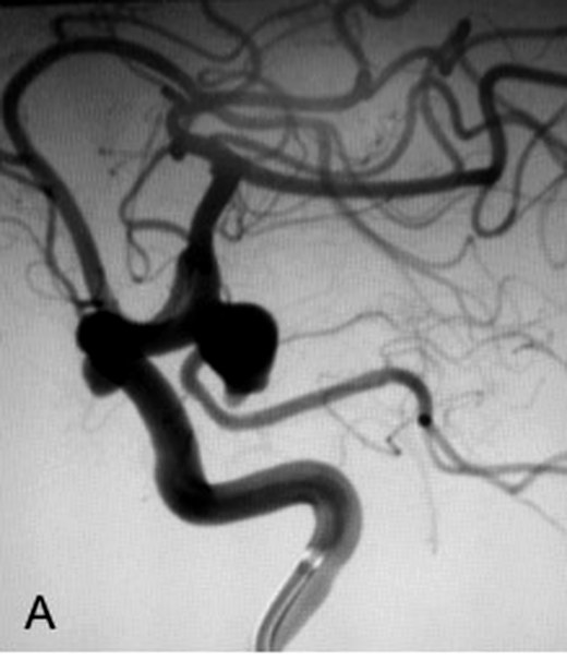
CT angiography or MR angiography usually finds an aneurysm and measures it. Catheter angiography (DSA) remains the most detailed view, showing the neck of the aneurysm, the branches arising near it and how blood flows around it — the details that decide which treatment is feasible. It is done under local anaesthesia through the wrist or groin, usually as a day procedure.
Treatment options
Endovascular coiling
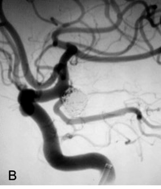
A catheter is guided from the wrist or groin into the aneurysm, and soft platinum coils are packed inside it until blood no longer enters. There is no incision on the head. Most patients go home within a day or two. Some aneurysms need a stent or balloon to hold the coils in place, which means taking blood-thinning medication for a period afterwards.
Flow diversion
Rather than filling the aneurysm, a fine braided stent is placed across its neck in the parent artery, redirecting flow past it so the aneurysm thromboses and shrinks over months. It has made some aneurysms treatable that previously were not — particularly wide-necked and large ones. It requires dual antiplatelet therapy and patience, since the result develops gradually.
Microsurgical clipping
Through a small opening in the skull, a titanium clip is placed across the neck of the aneurysm under the operating microscope. It remains the better answer for certain shapes and locations, particularly some middle cerebral artery aneurysms, and it gives an immediate and durable result. Hospital stay is longer and recovery slower than after a catheter procedure.
A surgeon who only clips will recommend clipping. A neurointerventionist who only coils will recommend coiling. Dr. Ambekar performs all three, which means the recommendation you receive is shaped by your anatomy rather than by the available tool.
If an aneurysm has ruptured
A ruptured aneurysm causes a subarachnoid haemorrhage — the sudden, worst-ever headache, often with vomiting, neck stiffness or loss of consciousness. It is a true emergency: the aneurysm must be secured quickly to prevent a second bleed, and the following two weeks require intensive monitoring for vasospasm and hydrocephalus. Treatment is either coiling or clipping, decided on the anatomy and the patient’s condition.
Do not wait to see whether it settles. Call +91 76780 63385 and dial 108 for an ambulance.
Questions worth asking at your consultation
- What is my actual annual risk of rupture, in numbers?
- What is the risk of the procedure you are recommending, in your hands?
- What happens if we simply watch, and how often would I be scanned?
- Will I need blood thinners afterwards, and for how long?
- Should my brothers, sisters or children be screened?