How an AVM shows itself
- Bleeding. The most serious presentation — sudden headache with weakness, speech difficulty or loss of consciousness.
- Seizures. A first seizure in adult life is a common way an AVM is discovered.
- Headache, sometimes long-standing and one-sided.
- Progressive neurological difficulty, as the malformation steals blood from surrounding brain.
- By chance, on a scan done for an unrelated reason.
Investigation
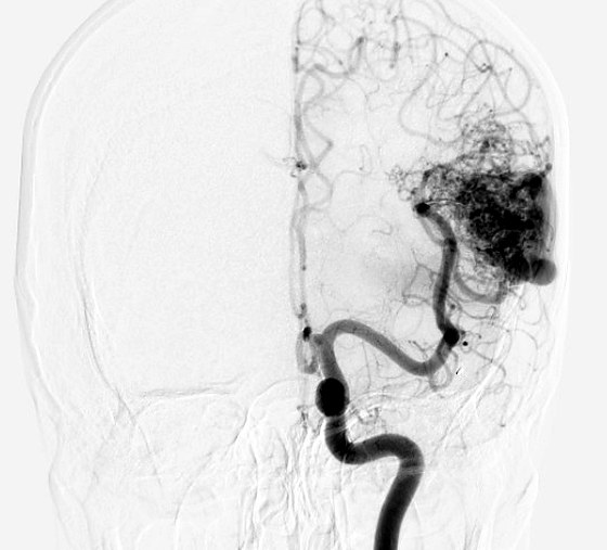
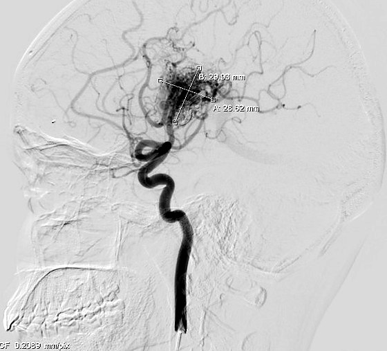
MRI shows where the malformation sits and its relationship to functional brain. Catheter angiography is essential before any treatment decision: it maps the feeding arteries, the nidus itself, the draining veins, and any associated aneurysms that raise bleeding risk. Functional MRI or tractography may be added when the AVM lies near speech or motor areas.
Treatment options
Embolisation
Liquid embolic material is injected through a microcatheter to close feeding vessels and the nidus. For some small AVMs this alone is curative. More often it is used to shrink the malformation and reduce blood loss before surgery, or to close a high-risk feature such as an associated aneurysm.
Microsurgical excision
Complete removal under the operating microscope, which gives an immediate and definitive cure. It is the treatment of choice for accessible AVMs, particularly after a bleed has already occurred.
Stereotactic radiosurgery
Focused radiation delivered in a single session causes the malformation to close over two to three years. It suits small, deep AVMs that would be dangerous to reach. The drawback is the waiting period, during which the risk of bleeding continues.
Combination and staging
Large AVMs are frequently treated in planned stages — embolisation followed by surgery, or embolisation followed by radiosurgery — rather than in a single sitting.
For an AVM that has never bled and sits in an area where treatment carries substantial risk, careful observation can be the safer course. Evidence from trials of unruptured AVMs has made the profession more cautious about intervening simply because a malformation exists. That conversation is worth having properly, with the angiogram on the screen.
What recovery looks like
After embolisation alone, most patients are home within a day or two. After open surgery, expect several days in hospital and a few weeks before returning to normal activity, longer if there has been a bleed. Seizure medication is often continued for a period. Follow-up angiography confirms that the malformation has been fully obliterated, because a partially treated AVM still carries risk.