Why this one matters so much
A spinal dural arteriovenous fistula raises the pressure in the veins draining the spinal cord. The cord becomes congested and swollen, and its function fails slowly — over months to years. Patients are investigated for lumbar canal stenosis, treated with physiotherapy, sometimes operated on for degenerative disease, while the real problem continues.
Closing the fistula stops the deterioration and often produces some recovery. But function already lost may not return. That makes early diagnosis worth far more than surgical skill here.
The pattern to recognise
- Gradually worsening weakness and heaviness in both legs, typically in a man over fifty.
- Numbness that begins in the feet and ascends.
- Symptoms worse after walking, standing or exertion — sometimes worse after a hot bath.
- Bladder urgency, hesitancy or incontinence appearing relatively early.
- Back pain that does not explain the neurological findings, and imaging of the spine that does not match the severity of the disability.
Investigation
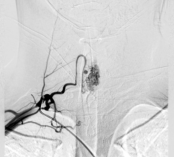
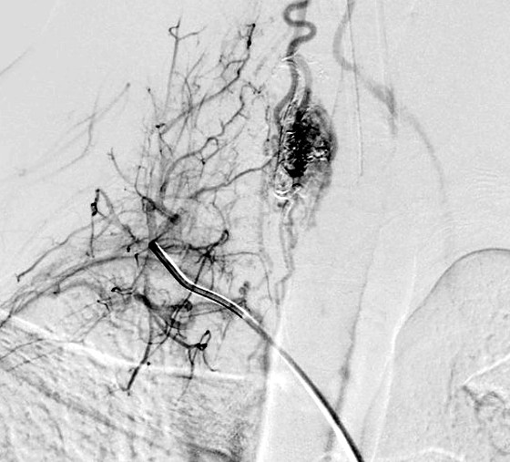
MRI of the whole spine shows swelling and signal change within the cord, often with dilated vessels visible on its surface — the finding that should trigger vascular imaging. Spinal angiography identifies the exact level and feeding vessel of the fistula. It is a meticulous study, since the fistula may lie several levels away from the cord changes it is causing.
Treatment
The fistula is closed either by embolisation through a catheter or by open surgical disconnection, which for many spinal dural fistulas is a small, well-tolerated and highly reliable operation. The choice depends on the anatomy and on whether the feeding vessel also supplies the cord.
Higher-flow spinal arteriovenous malformations and juvenile lesions are less common and more complex, and are often treated in stages.
Patients treated while still walking usually stabilise and often improve. Those treated after they have lost the ability to walk or lost bladder control frequently do not regain it. If the pattern above fits, ask for the MRI to be reviewed for vascular changes rather than only for disc disease.