How it presents
- A pulsing or whooshing noise in one ear, in time with the heartbeat. Frequently managed as an ear problem for months or years before anyone images the vessels.
- Headache, sometimes with raised pressure inside the head.
- Visual disturbance or eye symptoms, depending on which venous channels are involved.
- Haemorrhage or seizure, when the fistula drains backwards into the veins on the surface of the brain.
- Progressive confusion or unsteadiness, when venous drainage of the brain is obstructed.
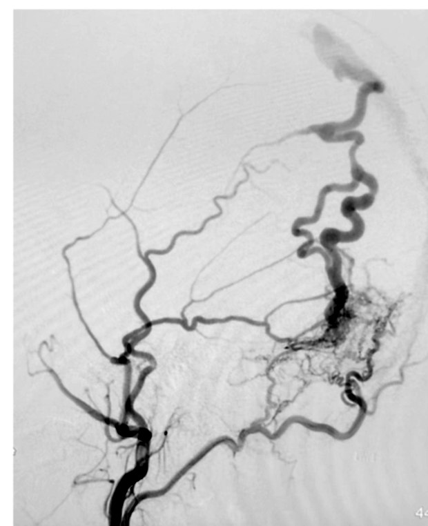
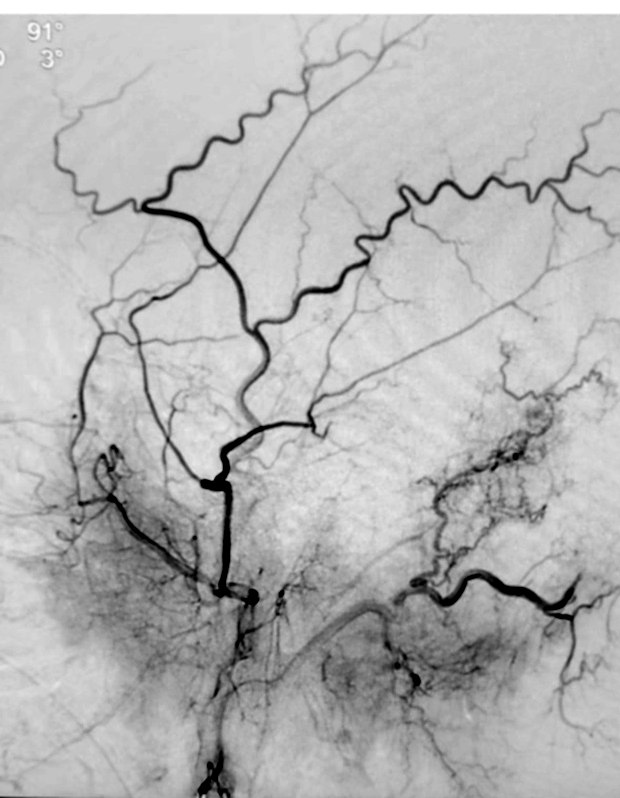
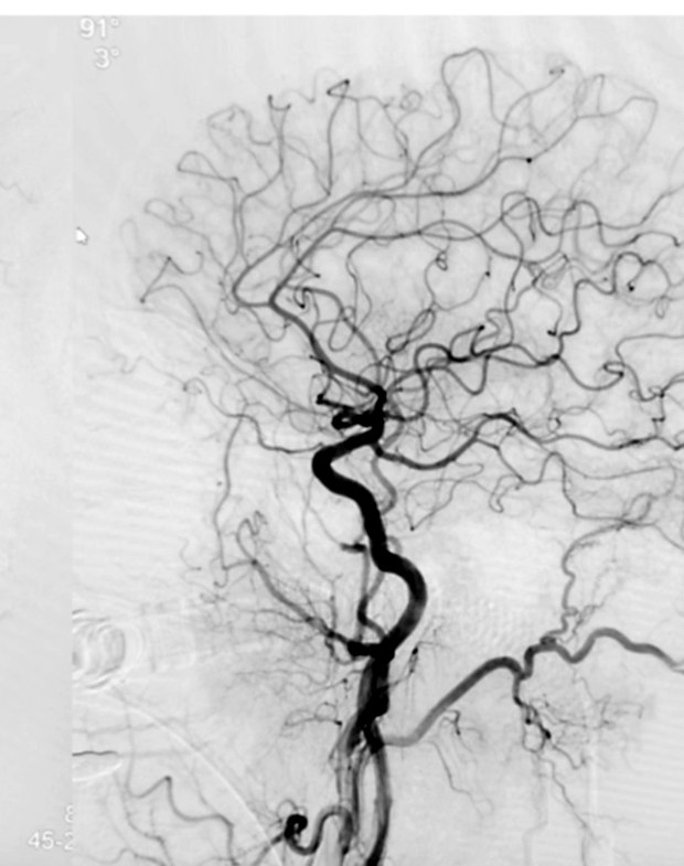
Why the drainage pattern matters more than the fistula
Not all dural fistulas are equally dangerous. What determines risk is where the blood goes. A fistula that drains forward into a normal venous sinus is often benign and may be treated only for symptoms. A fistula that drains backwards into cortical veins — vessels never designed for arterial pressure — carries a genuine annual risk of haemorrhage and generally needs closing regardless of how mild the symptoms are.
Establishing which pattern is present is the single most important thing the investigation must achieve.
Investigation
MRI and MR angiography may raise the suspicion, but they miss fistulas, and a normal MRI does not exclude one. Catheter angiography is the definitive test: it shows the feeding arteries, the exact point of the fistula, and above all the direction of venous drainage. When a pulsatile noise is convincing, a normal non-invasive scan is not a reason to stop looking.
Treatment
Most dural fistulas are closed endovascularly, in a single session. Liquid embolic material is delivered through a microcatheter, either along the arterial feeders or through the vein, to seal the connection at its point. The pulsing noise typically disappears immediately.
A small number require open surgical disconnection of the draining vein, and some benign fistulas that cause only a tolerable noise can reasonably be left alone once a dangerous drainage pattern has been excluded.
A noise that keeps time with your pulse, that changes when you press on your neck, or that stops when you turn your head, is a vascular sound until proved otherwise. It deserves a vascular investigation rather than another course of ear drops.